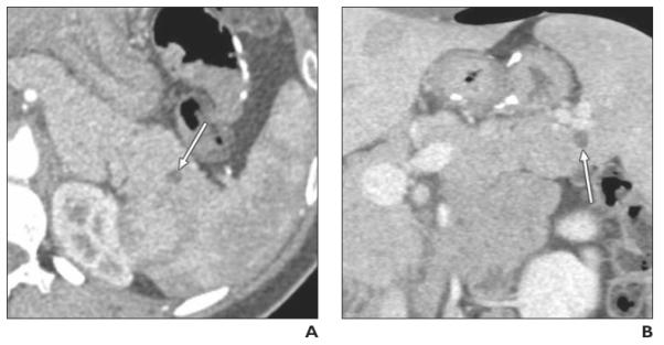
Fig. 1.

Well-differentiated pancreatic neuroendocrine tumor (NET), nonfunctioning, found on CT performed at another institution in 24-year-old woman with abdominal pain (case 3 in Table 2). Endoscopic ultrasound and fine-needle aspiration showed pancreatic NET.
A and B, Axial arterial phase (A) and coronal venous phase (B) multiplanar reformation images show purely cystic, unilocular cystic lesion (arrow) in tail of pancreas. CT differential diagnosis included intraductal papillary mucinous neoplasm and other cystic lesions.
