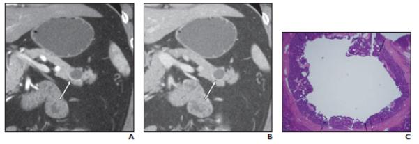
Fig. 2.

Well-differentiated neuroendocrine tumor (NET), nonfunctioning, in 56-year-old man. Cystic mass was initially found on CT performed for abdominal pain; mass had increased in size during follow-up. Fine-needle aspiration performed at another institution revealed well-differentiated NET (case 2 in Table 2).
A,Coronal arterial phase multiplanar reformation (MPR) image shows purely cystic mass in tail of pancreas with equivocal minimal smooth rim of enhancement along inferior border (arrow).
B,Coronal venous phase MPR image shows purely cystic mass (arrow) without detectable peripheral enhancement.
C,Photomicrograph (H and E, 2×) shows there is unilocular cyst in center of tumor. Cyst is lined by neuroendocrine cells.
