
Fig. 3.
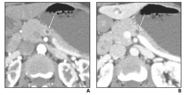
Well-differentiated neuroendocrine tumor (NET), nonfunctioning, in 47-year-old woman. Small cystic lesion was incidentally found on CT performed for evaluation of hematuria. Endoscopic ultrasound and fine-needle aspiration revealed pancreatic NET (case 6 in Table 2).
A,Axial arterial phase image shows partially (> 50%) cystic mass with thin, smooth peripheral enhancement (arrow) greater than that of pancreatic parenchyma.
B,Axial venous phase image shows small cystic mass (arrow) with no detectable peripheral enhancement.