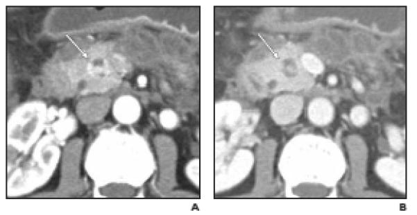
Fig. 5.

Well-differentiated neuroendocrine tumor (NET), nonfunctioning, in 51-year-old woman. Pancreatic head mass was found on CT performed for evaluation of pancreatitis (case 14 in Table 2).
A and B,Arterial phase (A) and venous phase (B) axial images show partially (≤50%) cystic small mass in head of pancreas (arrow). Note that intense contrast enhancement of peripheral and internal nodular components is visible only on arterial phase. Peripancreatic inflammation and fluid collections are due to pancreatitis. CT differential diagnosis included pancreatic NET and other tumors.
