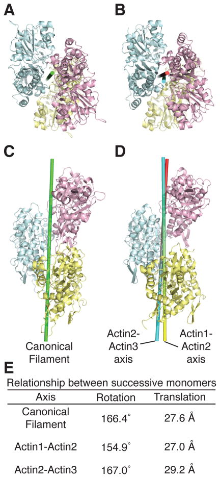
Figure 4. Actins in the VopL complex resemble the canonical actin filament. See also Figure S4.
(A and C) Canonical actin filament (Oda et al., 2009).
(B and D) Actins from the VopL complex, colored as in Fig. 1. Axes relating pairs of actin monomers are shown as cylinders.
(E) Rotations and translations associated with the depicted axes.

