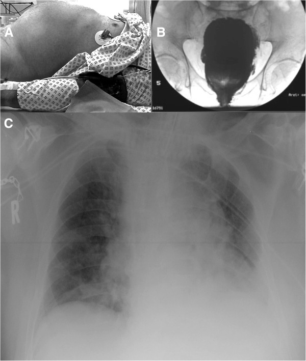
Figure 1.

Patient images. A) Patient on the x-ray table. Please note the prominent abdominal region. Measured intra-abdominal pressure was 26 mmHg. B) Contrast agent x-ray of the bladder. There are no signs of bladder perforation. C) X-ray of the patient’s thorax during ventilation. Endotracheal tube and central venous line were in the correct position. There were signs of atelectasis of the left lower lung and cephalic displacement of the diaphragm due to the intra-abdominal volume augmentation and despite the restricted conditions for the x-ray in the ICU bed.
