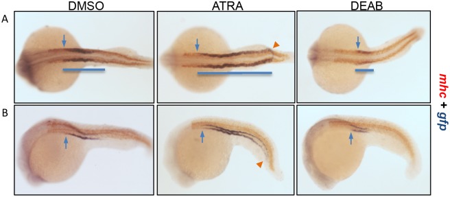
Figure 8. RA signaling regulates pronephric tubule segmentation.

The Tg(gtshβ::GFP) embryos at bud stage were treated with DMSO (as control), ATRA or DEAB, and fixed at 22 hpf to perform double whole-mount in situ hybridization with gfp (purple) and mhc (red) probes. (A) Dorsal view. (B) Lateral view. Cloacas are indicated by arrowheads. The most anterior positions of gfp expression are indicated by arrows. The blue lines are drawn to mark the gfp expression patterns.
