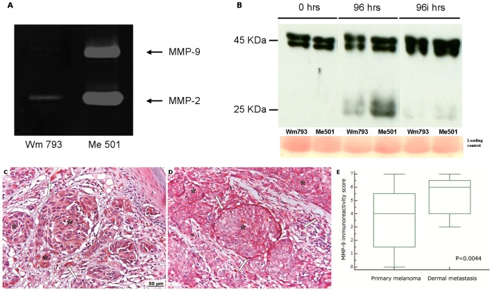
Figure 2. Different expression of MMP-9 in primary and metastatic melanoma.
(A) Detection of MMP-2 and MMP-9 activity in the culture media of WM793 and Me501 cells by zymographic assay. The arrows point to the areas of degradation of the gelatine matrix produced by the indicated proteases (B) Western blot analysis showing degradation over time of IGFBP-3 by melanoma-produced proteases. Human recombinant IGFBP-3 was incubated for the indicated times with the culture media of the primary melanoma line WM793 or of the metastatic line Me501, in the absence (96 hrs) or in the presence (96i hrs) of protease inhibitors. A Red Ponceau staining of the original gel is shown on the bottom as the loading control. (C,D) MMP-9 immunostaining of primary (C) and metastatic (D) melanoma. Asterisks indicate melanocytes nests and the arrows indicate stromal cells. Original magnification, X100. (E) Comparison of the MMP-9 reactivity score of primary and metastatic melanoma. Central boxes represent values from lower to upper quartile (25th–75th percentile). Middle lines represent median. Vertical lines extend from minimum to maximum value. P value was calculated by Mann-Whitney U test. The reactive cells were counted, and the scores determined, as described in Fig. 1 and in the Methods section.

