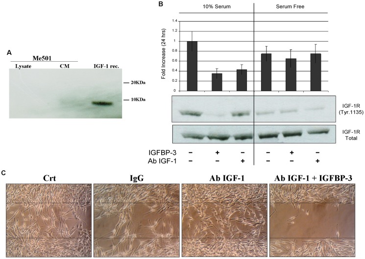
Figure 6. IGFBP-3 acts independently of IGF-1.
A) Western blot analysis for the detection of IGF-1 in lysates or culture media (CM) of Me501 cells. IGF-1 rec represents the positive control with recombinant IGF-1 (5 ng). B) Western blot analysis for detection of Tyr 1135-phosphorylation on the IGF-1 receptor β. Me501 cells were grown for 24 h in the presence of 10% serum (left panels) or in the absence of serum (right panels). For each group, treatments with IGFBP-3 or with anti-IGF-1 antibodies were performed. The amount of phosphorylated IGF-1-β receptor was quantified by densitometric analysis using total receptor (which remained unchanged) as the internal standard. C) Scratch-repair capacity of Me501 cells in the presence of anti-IGF1-antibodies and of IGFBP-3. The images shown are representative of experiments that were repeated at least three times.

