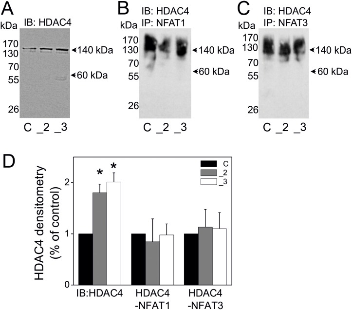
Figure 4. The interaction of NFAT1 and NFAT3 with HDAC4 isoform in PMCA2- or PMCA3-deficient PC12 cells.
RIPA-total cellular extracts were subjected to immunoblotting to verify HDAC4 protein content and served as inputs of immunoprecipitation (A). The cellular extracts (inputs) were incubated with protein A/G agarose beads and with anti-NFAT1 antibody (B) or with anti-NFAT3 antibody (C) and the obtained immunoprecipitates were subjected to immunoblotting for HDAC4. All immunoblots and immunoprecipitates were measured densitometrically and expressed as % of control cells (D). Student’s t-test was used for comparison of control cells with PMCA2- or PMCA3- deficient cells. *P≤0.05, n = 3. Symbols: control cells (C), PMCA2-deficient cells (_2), PMCA3-deficient cells (_3).

