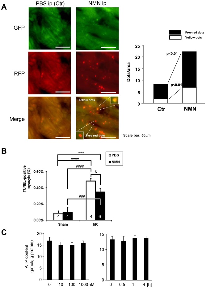
Figure 7. NMN stimulates autophagy in control hearts.
A, Either NMN (500 mg/kg per injection) or vehicle (PBS) was administered (i.p. injection) to Tg-mRFP-GFP-LC3 mice. After 2 hours, the number of fluorescent LC3 dots was evaluated. Representative images of GFP puncta, mRFP puncta and their merged images are shown. The results of the quantitative analysis of RFP only dots and RFP/GFP double positive (yellow) dots/area are also shown. B, Either NMN (500 mg/kg per injection) or vehicle (PBS) was administered (i.p. injection) to mice 30 minutes before I/R, and then the mice were subjected to either 30 minutes ischemia followed by 24 hours of reperfusion or sham operation. The extent of cardiomyocyte apoptosis in the border zone was evaluated with TUNEL staining. The results of the quantitative analysis of TUNEL-positive cardiomyocytes are shown. C. Three days after isolation, cardiomyocytes were treated with the indicated dosage of NMN for 30 minutes (Left) and with 1000 nM NMN for the indicated time (Right). ATP contents were measured by ATP Bioluminescent Assay Kit (Sigma). n = 4 (Left) and 3(Right).

