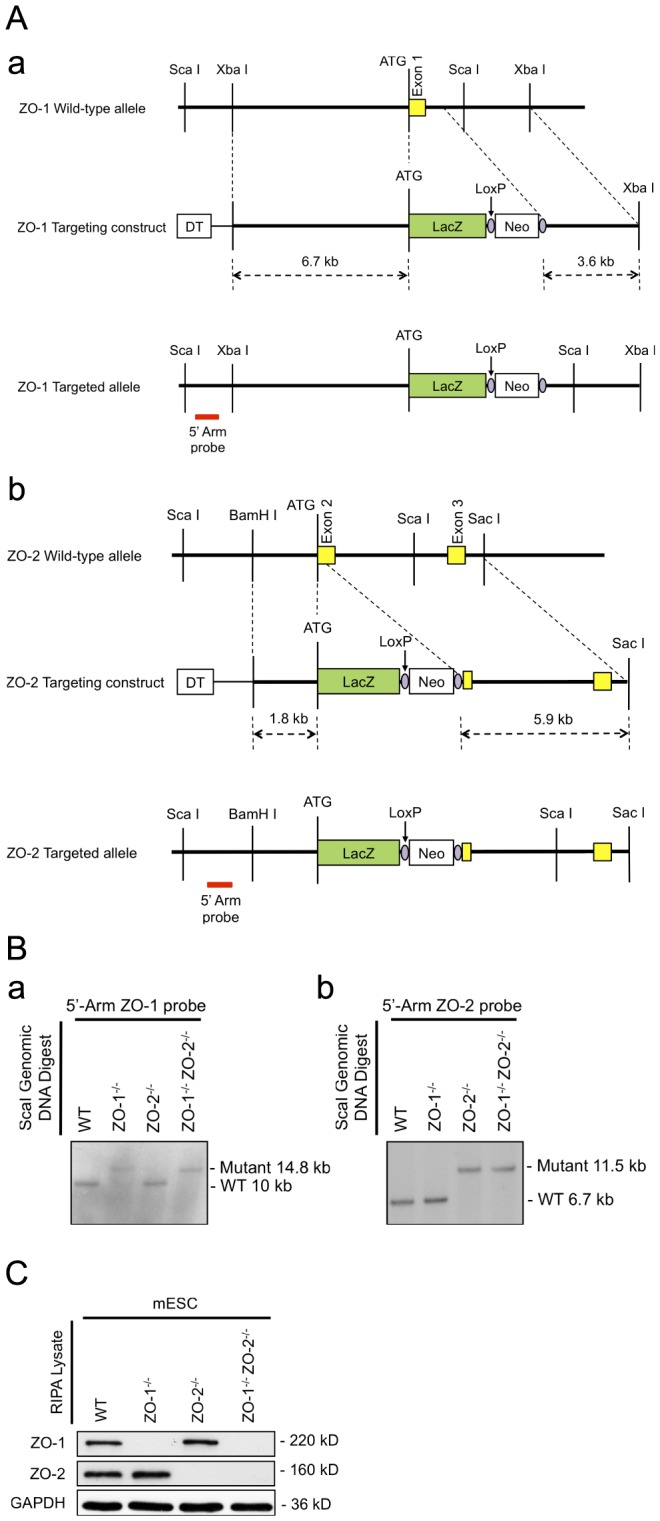
Figure 1. Targeting of the ZO-1 and ZO-2 locus and genotyping.

(A) Targeting strategy. Schematic representation of the genomic loci restriction maps of ZO-1 showing exon 1 (panel a) and ZO-2 showing exons 2–3 (panel b) in yellow boxes with the initiation ATG. Through the in-frame insertion of a LacZ gene (green box) and a loxP-flanked (purple circle) Neo cassette (white box) immediately downstream of the ATG codon, the ZO-1 and ZO-2 allele-targeting constructs were designed to delete the entire ZO-1 exon 1 and part of the downstream intron; and part of ZO-2 exon 2 respectively. The red bar indicates the position of probe hybridization for Southern blot analysis. (B) Genotypic analysis by Southern blotting. ScaI-digested genomic DNA of selected mESC clones were hybridized with a DIG-labelled 5′ genomic DNA probe for the identification of homologous recombinants. 10 kb and 14.8 kb probe-hybridized fragments correspond to WT and targeted allele of ZO-1 locus respectively (panel a), whereas WT and targeted allele of ZO-2 locus are respectively represented by 6.7 kb and 11.5 kb fragments (panel b). (C) Protein expression analysis. mESC lysates were subjected to immunoblotting with anti-ZO-1 and anti-ZO-2 antibodies. The presence of ZO-1 and ZO-2 protein is indicated by 220 kD and 160 kD bands respectively. GAPDH served as a control for equal lysate input.
