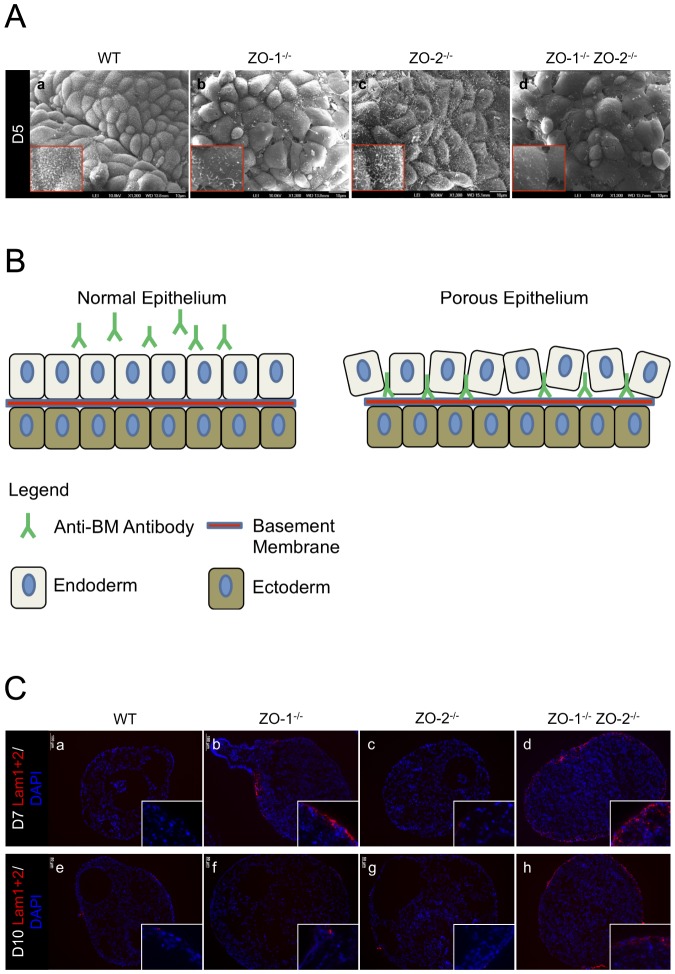
Figure 3. ExEn organization and permeability is abnormal in EBs lacking ZO-1 and ZO-2.
(A) Scanning electron micrographs. ExEn apical surface morphology of WT control and ZO gene-deleted EBs was analyzed by SEM. The apical surface of compacted ExEn cells can be seen covered with dense hair-like microvilli in WT (panel a) and ZO-2-/- (panel c) EBs. ZO-1-/- (panel b) and ZO-1-/- ZO-2-/- (panel d) EB ExEn cells are more loosely packed and bear sparse microvilli formation. Magnification of image in insets. (B) ExEn permeability assay illustration. Intact ExEn of live EBs pre-incubated with anti-BM IgG will not allow penetration of the antibodies to the underlying BM. In a permeability-compromised ExEn, the anti-BM IgG can move across the ExEn paracellularly and bind to the underlying BM. (C) Permeability assay using anti-Lam1+2 pre-incubate. Live WT, ZO-1-/-, ZO-2-/- and ZO-1-/- ZO-2-/- EBs at Day-7 (panels a-d) and Day-10 (panels e–h) of culture were pre-incubated for 2 hrs in medium spiked with IgG recognizing the BM component Laminin1+2. The EBs were subsequently washed and fixed, and the embedded sections were then permeabilized and treated with fluorophore-tagged secondary antibodies to visualize the anti-Lam1+2 IgG (red color). Nuclei are labeled with DAPI (blue color). Magnification of image in insets.

