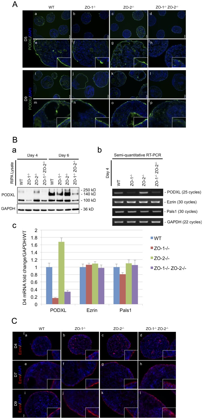
Figure 5. PODXL expression and Ezrin localization at the ExEn is aberrant when ZO-1 and ZO-2 is deleted.
(A) Immunofluorescence staining of PODXL. EB cryosections from Day-5 (panels a–h) and -9 (panels i–p) cultures were immunostained for PODXL (green color). Nuclei are labeled with DAPI (blue color). Magnification of image in insets. (B) PODXL expression. Protein expression of PODXL at Day-4 and -6 was determined by immunoblot. 100 (immature glycosylated), 140 (mature glycosylated) and 250 (dimer) kD bands represent the various post-translationally modified forms of PODXL. GAPDH was used as a lysate loading control (panel a). PODXL transcript level was analyzed by semi-quantitative RT-PCR. Reverse transcribed cDNA was amplified with specific primer sets at optimized cycle numbers (indicated on right side of panels). Ezrin and Pals1 were selected as epithelia polarized controls. GAPDH amplification served as a control for equal RNA input (panel b). Quantitative real-time PCR was also employed to validate PODXL transcript expression. Expression levels are presented as average fold-change of three separate experiments normalized to GAPDH and relative to WT control (panel c). (C) Immunofluorescence staining of Ezrin. Cryosections of EBs harvested at Day-4 (panels a–d), Day-7 (panels e–h) and Day-9 (panels i–l) of culture were immunostained with antibodies against Ezrin (red color). Nuclei are labeled with DAPI (blue color). Magnification of image in insets.

