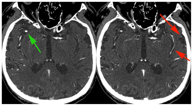
Figure 3. Example of delayed collateral vessels filling beyond a proximal occlusion.
83 year old male with atrial fibrillation and acute stroke. Early phase images show occlusion of the proximal M1 segment of the left MCA and robust opacification of the anterior division of the right MCA (green arrow). Delayed phase images show opacification of several pial vessels over the left temporal convexity and in the left Sylvian fissure (red arrows), delayed because of pial-pial collateral flow beyond the occlusion. Note that normal vessels demonstrate reduced opacification compared with the initial phase images (green arrow); this is due to decreased contrast concentration as a result of recirculation of the initial bolus. This CTA was obtained after MRI demonstrated the extent of MCA territory infarction; no management decisions were changed as a result of the delayed views.

