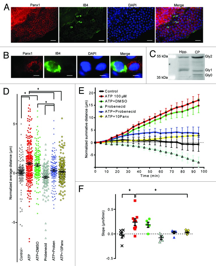
Figure 3. Panx1 channels are involved in epiplexus cells activation by exogenous 100 µM ATP. Panx1 channels are robustly expressed on choroidal epithelium, but were rarely detected in epiplexus cells. Western blotting analysis confirmed presence of Panx1 in the CP. The Panx1 blockers, 500 µM probenecid and 100 µM 10panx significantly decreased ATP-triggered chemokinesis. (A and B) Immunofluorescent staining for Panx1 in the CP and on an individual IB4-positive epiplexus cell. (C) Detection of Panx1 protein by western blotting. (D) Normalized average distance and statistical analysis, each symbol represents a single cell and all cells from the experiments are shown; (E) normalized summative distance; (F) slope of normalized summative distance where each symbol represents a single isolated CP. Scale bars are 50 μm (A) and 5 μm (B).

An official website of the United States government
Here's how you know
Official websites use .gov
A
.gov website belongs to an official
government organization in the United States.
Secure .gov websites use HTTPS
A lock (
) or https:// means you've safely
connected to the .gov website. Share sensitive
information only on official, secure websites.
