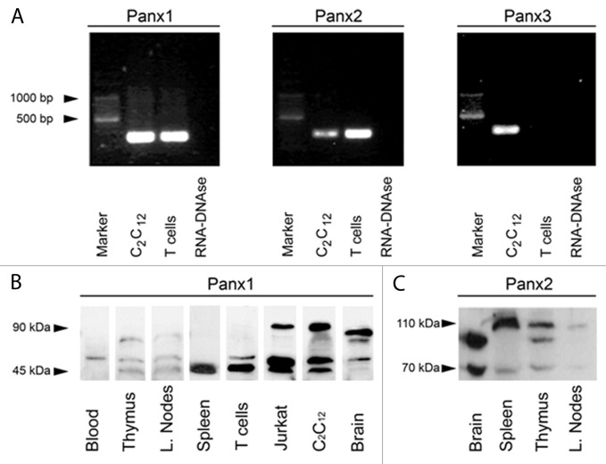
Figure 1. Panxs 1 and 2 are present in mouse T cells and secondary lymphoid organs. (A) RT-PCR products generated with specific primers for Panx1, Panx2 and Panx3 were obtained from RNA extracts of T cells isolated from lymph nodes of Balb/c mice and C2C12 cells used as positive control. Panx1 amplicon was detected from RNA purified from T cells and C2C12 cells at the expected electrophoretic mobility (∼280 bp). Panx2 amplicon was also present in both samples (∼270 bp). However, Panx3 amplicon was detected in C2C12 cells but not in T cells. No amplicons were detected in samples having RNA-DNase or water (not shown) that were used as DNA-contamination controls for mRNA isolation and PCR amplification control, respectively. (B) Immunoblot of Panx1 in total homogenates of mouse blood, thymus, axillar lymph nodes and spleen, as well as freshly isolated T cells obtained from lymph nodes of BALB/c mice and Jurkat cells. C2C12 cells and mouse brain were used as positive controls. One to four main bands reactive to anti-Panx1 antibody were detected at 45–50 kDa and 90 kDa. (C) Immunoblot analysis of Panx2 in total homogenates of spleen, thymus and lymph node. Brain was used as Panx2 positive control. Panx2 reactive bands were detected mainly between 90–110 kDa.

An official website of the United States government
Here's how you know
Official websites use .gov
A
.gov website belongs to an official
government organization in the United States.
Secure .gov websites use HTTPS
A lock (
) or https:// means you've safely
connected to the .gov website. Share sensitive
information only on official, secure websites.
