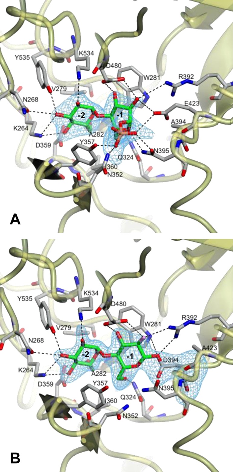
Figure 2.

Structures of GlgE with (A) α-maltose 1-phosphate bound to the D394A mutein (PDB entry 4CN1) and (B) the covalent intermediate formed between the E423A mutein and 2-deoxy-2-fluoro-α-maltosyl fluoride (PDB entry 4CN4). Difference electron density “omit” maps were generated for bound ligands using phases from final models without ligand coordinates after application of small random shifts to the models and re-refining. The corresponding stereo images are shown in Figure S1 of the Supporting Information. Some amino acids interacting with the ligands have been omitted for the sake of clarity, but all are shown in Figure S2 of the Supporting Information. Subsites −1 and −2 are labeled.
