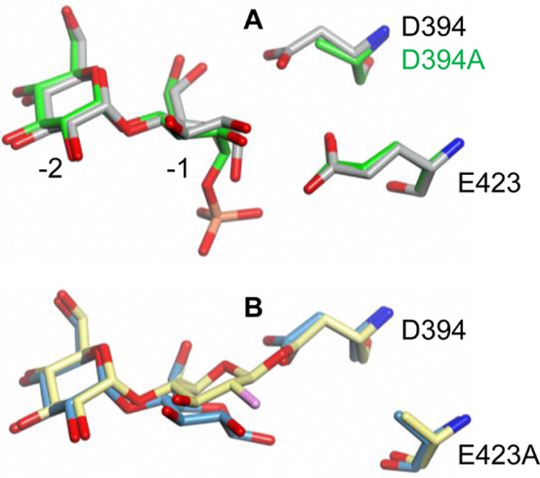
Figure 3.

Superposed structures of (A) maltose (PDB entry 3ZT5, gray) and α-maltose 1-phosphate (PDB entry 4CN1, green) bound to wild-type and D394A GlgE, respectively, and (B) maltose (PDB entry 4CN6, blue) and the trapped 2-deoxy-2-fluoro-β-maltosyl intermediate (PDB entry 4CN4, yellow) bound to the E423A mutein. Comparison of the structures in panel B reveals that the anomeric carbon moves 1.6 Ǻ to allow the formation of a covalent bond between the disaccharide and D394. The glucose rings in each structure adopt the low-energy 4C1 conformation. The orientation shown is similar to that in Scheme 1 with subsites −1 and −2 labeled in panel A.
