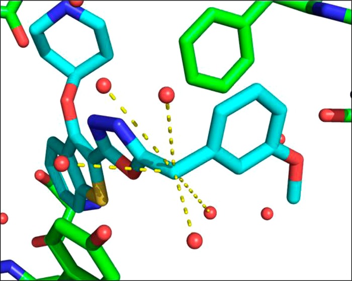
Figure 3.

X-ray crystal structure of 20b (blue) bound to PvNMT (green). Further inspection of the water molecules within the active site shows that the benzylic CH2 occupies a heavily solvated pocket, indicating that substitution may result in more favorable energetics within the enzyme active site. Dashed lines indicate water molecules within 5 Å of the benzylic position.
