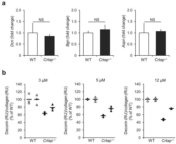
Figure 3.
Reduced decorin binding to type I collagen of Crtap−/− mice. (a) Quantitative RTPCR of calvarial bone of P3 mice for the small leucine-rich proteoglycans decorin (Dcn), biglycan (Bgn), and asporin (Aspn) in WT and Crtap−/− mice. Results given as fold change of the mean of WT group±SD, n=5 per group. NS, not significant. (b) Surface plasmon resonance analysis measuring the binding of recombinant decorin core protein to type I collagen of WT and Crtap−/− mice. Three technical replicates at each of the indicated concentrations of decorin were performed in two independent biological replicates (◆ replicate 1, ▲ replicate 2). Results are shown as the percentage of the mean of WT (bars indicate mean per group).

