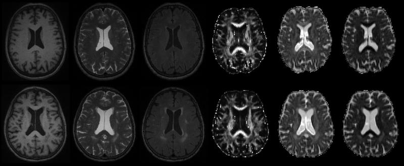
Figure 2.
Multiple image types in an older adult without (top) and with (bottom) white matter lesions. Variation in periventricular lesion contrast is obvious across the different image modalities, and this variation may provide important quantitative information allowing the specification of lesion pathology. In order from left to right: T1, T2, FLAIR, diffusion tensor imaging (DTI) fractional anisotropy, DTI axial diffusivity, DTI radial diffusivity.

