
Figure 5.
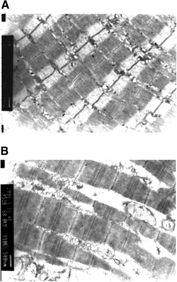
Electron microscopy. (A) Normal skeletal muscle obtained after severing the rat limb but before freezing. Regularly arranged myofibrils, clear z-lines, dense mitochondrial cristae, and intact mitochondrial membranes without swelling were seen. (B) Skeletal muscle of a failed replantation after cryopreservation. Images were obtained four hours after reperfusion. Some myofibrils were ruptured with unclear z-lines, the mitochondria exhibited maximal swelling, crests were disarrayed, and parts of the myofibrils showed vacuolation.