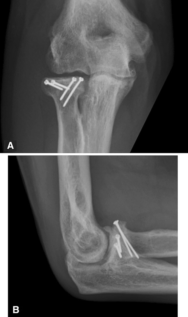
Fig. 3A–B.

(A) AP and (B) lateral radiographs of the patient shown in Figure 2 taken 4 years postoperatively after interim capsular release and coronoid hardware removal demonstrate progression of arthrosis despite maintained alignment. Note the capitellar osteopenia, subchondral bone irregularity, and ulnotrochlear degenerative changes.
