
Fig. 1.
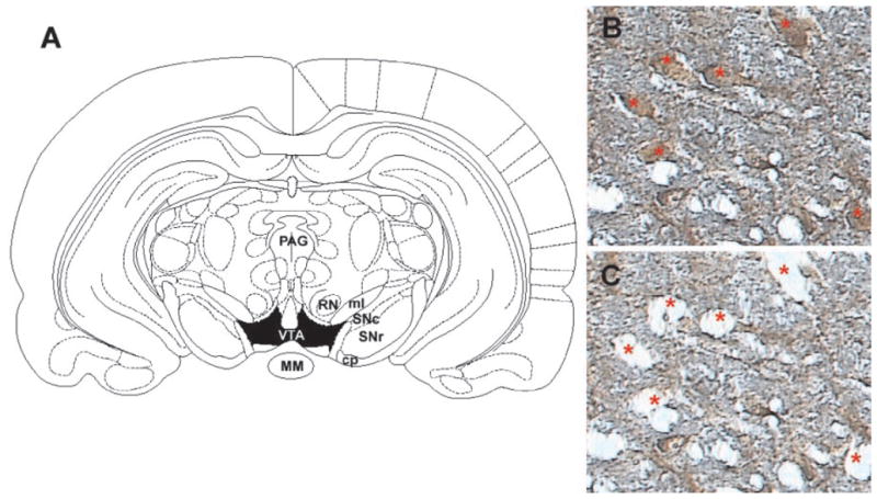
A, schematic of region for laser capture microdissection. Rat coronal midbrain section at −5.30 mm (relative to bregma) showing boundaries for VTA (shaded in black) from which tyrosine hydroxylase immunopositive cells were dissected. Midbrain section immunolabeled with anti-TH antibody (B) and the same section after microdissection of the indicated neurons (C) under 20× magnification. Note the specificity of the dissections and the minimal disruption of surrounding neuropil. MM, mammillary nucleus; PAG, periaqueductal gray area; SNc, substantia nigra-pars compacta; SNr, substantia nigra-pars reticulata; RN, red nucleus; ml, medial lemniscus; cp, cerebral peduncle.