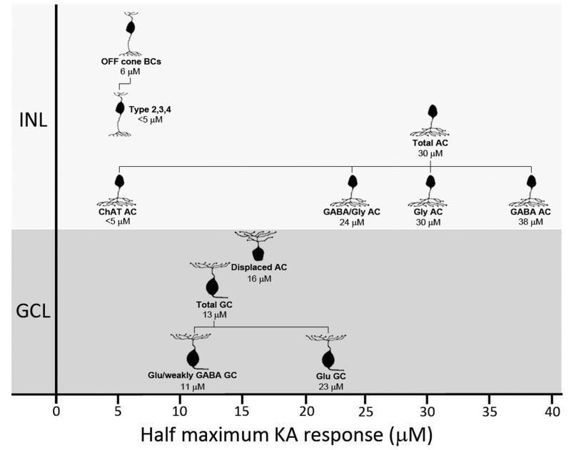
Figure 13.
Diagrammatic representation of kainate (KA) sensitivity of various neurochemically identified cell populations in the rat inner retina. Annotations below cell images indicate the neurochemical cell class and the half-maximal activation concentration for that class. Data are based on dose activation curves in Figures 3, 10, and 12. For Chat ACs and type 2, 3, and 4 BCs, estimates of half-maximum concentration have been made from immunolabeling. Abbreviations: INL, inner nuclear layer; GCL, ganglion cell layer; BC, bipolar cell; AC, amacrine cell; GC, ganglion cell; GABA, γ-aminobutyric acid; Gly, glycine; Glu, glutamate.

