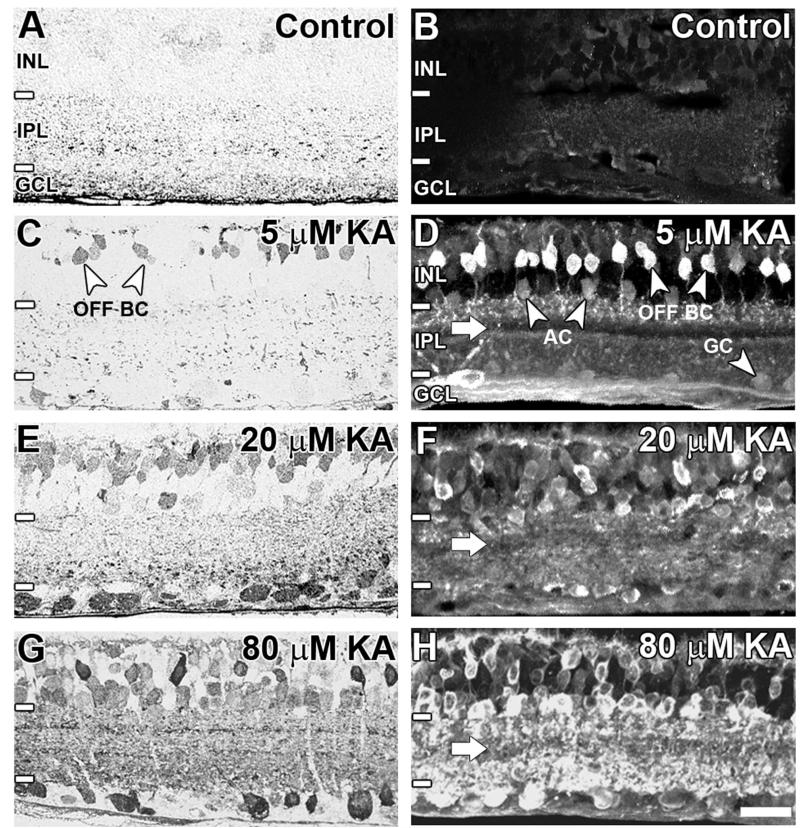
Figure 2.
AGB immunoreactivity in the adult rat retina at (A, B) basal levels, (C,D) 5 μM kainate (KA) activation, (E,F) 20 μM KA activation, and (G,H) 80 μM KA activation. Sections were imaged with brightfield light microscopy using postembedding immunocytochemistry (A,C,E,G) or confocal microscopy using indirect immunofluorescence (B,D,F,H). Retinal layers including the inner nuclear layer (INL), inner plexiform layer (IPL), and ganglion cell layer (GCL) are indicated by white lines and annotations on the left-hand side of the image. The white arrow indicates weakly AGB-immunoreactive strata in the IPL. BC, bipolar cell; AC, amacrine cell; GC, ganglion cell. Scale bar = 20 μm in H (applies to A-H).

