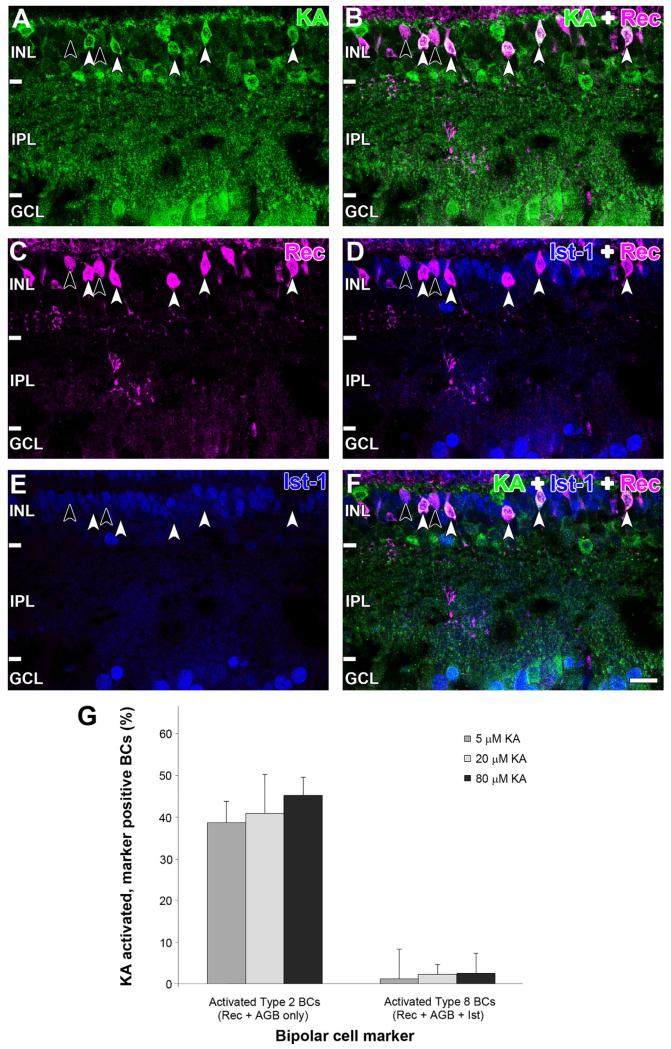
Figure 9.
Immunoreactivity of (A) AGB (green), (B) recoverin (magenta), and (C) islet-1 (blue) after activation with 80 μM kainate (KA). D: Overlap of AGB and recoverin channels shows that many recoverin BCs were activated by KA (white arrowheads) but not all (black arrow-heads). E: Overlap of recoverin and Islet-1 channels. Type 8 BCs are identified by colocalization of recoverin and Islet-1 (black arrow-heads), and type 2 are identified by the absence of Islet-1 staining (white arrowheads). F: Overlap of all three markers. Type 8 BCs are not activated by KA (black arrowheads), whereas type 2 BCs are activated by KA. Retinal layer annotations are the same as in Figure 2. G: Quantification of recoverin BC populations with functional KA receptors. The two cell types are presented as a percentage of the total recoverin BC population (5 μM: n = 735; 20 μM: n = 599; 80 μM: n = 455). Each column represents the mean and SD from five independent rat retinae. Abbreviations: Rec, recoverin; Ist-1, islet-1; BC, bipolar cell. Scale bar = 20 μm in F (applies to A–F).

