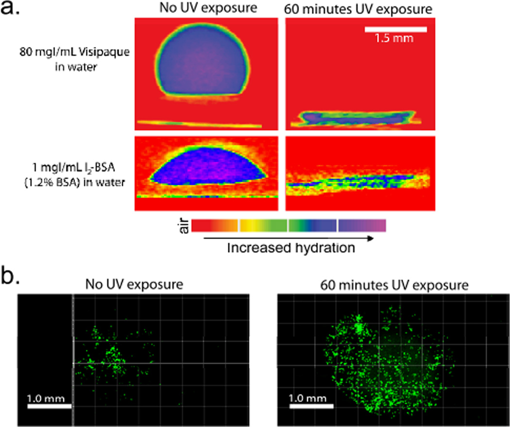
Figure 3.
(a) µCT imaging of 7:3 PCL:PGC-C12-NPE electrospun meshes exposed to either 0 minutes (left) or 60 minutes (right) of 365 nm UV light through a 1590 µm in diameter photo mask. Visipaque (left) in water or an I2-BSA solution (right) was used to track wetting/adsorption into the mesh. (b) Fluorescence images of MCF7 cells (green) seeded onto meshes exposed to 0 minutes (left) and 60 minutes (right) of UV light.

