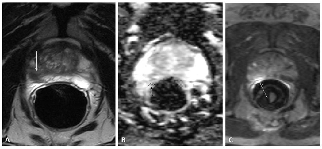
Figure 1. 70 year old patient with Gleason 3+3 prostate carcinoma.

A: T2 Weighted image, with hypodense lesion (arrow) B: Diffusion Weighted Image. The arrow indicates an area with diffusion restriction C: Dynamic Contrast Enhanced Image. The arrow indicates an enhancing lesion.
