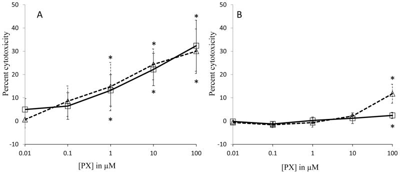
Fig. 1.
Cytotoxicity of paraoxon (PX) on HSG cells. Cells were treated with PX and analyzed at 24 h (squares) and 48 h (triangles). Cytotoxicity was estimated by a MTT colorimetric assay (A), and an LDH release assay (B). Results are presented as average with standard deviation compared to cells treated with vehicle (0.1% ethanol). Triton X-100 was used as a positive control for cell death (100% cytotoxicity). *A significant difference between the treatment and negative control (no PX) was determined (p < 0.05) using one-way ANOVA followed by a Tukey-Kramer post host test (n = 6–8).

