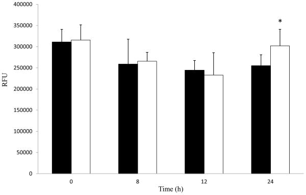
Fig. 2.

Paraoxon (PX) exposure slightly alters cellular ATP levels in HSG cells. Cells were treated with 0.1% ethanol as control (black bars) or 10 μM PX (white bars). Cellular lysates were analyzed for cellular ATP levels (RFU, relative fluorescence units) from 8 to 24 h. Samples were analyzed and results shown are averages with standard deviations. Samples were analyzed for significance with Student’s t-test, * represents p values < 0.05 (n = 4–6).
