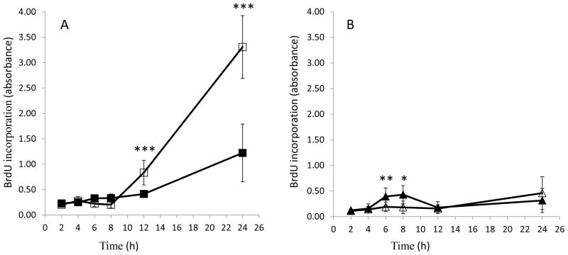
Fig. 3.
Paraoxon (PX) exposure induces DNA fragmentation in HSG cell lysates. Cell lysates (Panel A) or cell culture supernatants (Panel B) were analyzed for BrdU incorporation at 2 to 24 h following 10 μM PX treatment (or ethanol control). Black squares, control lysates; black triangles, control supernatants; white squares, PX lysates; white triangles, PX supernatants. Results were analyzed using Student’s t-test and data are represented as average with standard deviation. * represents p values < 0.05, ** represents p values < 0.01, and *** represents p values < 0.001 (n = 6–8).

