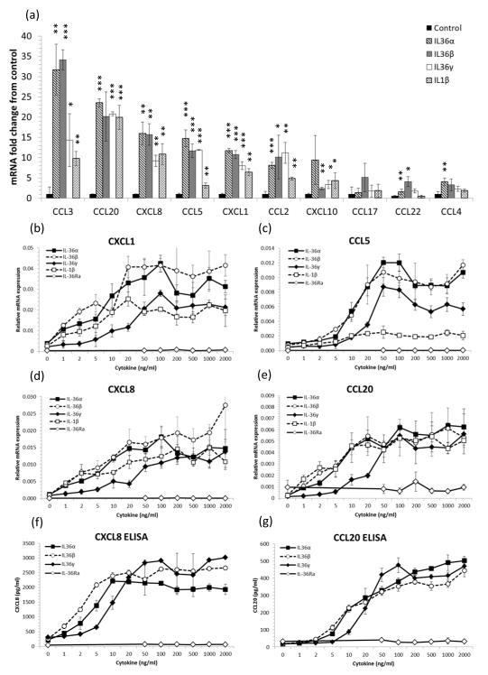
Figure 1. IL-36 cytokines induce chemokine expression by keratinocytes.
4-day post-confluent normal human keratinocytes (NHK) were stimulated for 24h with recombinant truncated IL-36R ligands or IL-1β. Total RNA was extracted and mRNA transcripts quantified by qRT-PCR relative to the housekeeping gene RPL-P0 and conditioned medium assayed by ELISA. 100ng/ml IL-36α, IL-36β and IL-36γ significantly induced T cell chemokine mRNA expression compared with untreated cells, mean ± S.D. (n=3) (a). IL-36α, IL-36β, IL-36γ and IL-1β but not IL-36Ra dose-dependently induced CXCL1, CCL5, CXCL8 and CCL20 mRNA expression (b-e) and CXCL8 and CCL20 protein secretion (f and g) by keratinocytes. Mean ± S.D. (n=3). Statistical significance indicated by * p<0.05, t** p<0.01 or *** p<0.001, Student’s t-test.

