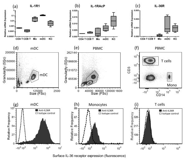
Figure 3. Human APC but not T cells express the IL-36 receptor.
Keratinocytes (KC), monocytes and myeloid DC (mDC) express IL-1R1, IL-1RAcP and IL-36R mRNA transcripts, however, CD4+ and CD8+ T cells were found not to express IL-36R as determined by qRT-PCR (a-c, n=4 donors). Flow cytometric analysis reveals that in contrast to mDC (d and g) and monocytes (e, f and h), T cells (e, f and i) did not express surface IL-36R. Filled histogram: anti-IL36R, dotted histogram, isotype control antibody. Flow cytometry gating shown in panels 3d-f; Flow cytometry data are representative of 6 donors.

