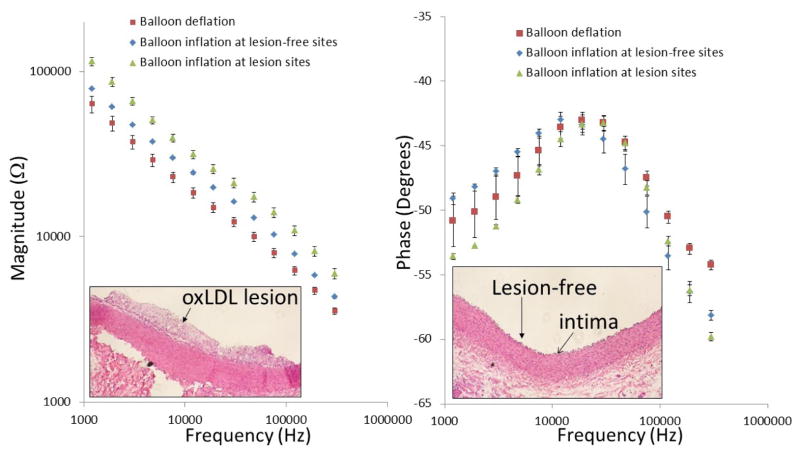
Fig. 4.
Ex-vivo EIS acquisition from explanted rabbit aorta and corresponding histology of the aorta wall. After 8 weeks of high-fat diet, rabbits were sacrificed and aortas were extracted and fixed in 15% paraformal aldehyde (PFA). The individual aortas were sectioned into 2–3cm segments and immersed in PBS for deployment of the balloon-inflatable EIS sensor. Endoluminal lipid deposition or atherosclerotic lesions were visible for EIS interrogation and comparison with the disease-free sites. (a) After the balloon inflation, the impedance magnitude increased over the entire frequency range from 1 kHz to 1,000 kHz. EIS measurement further increased significantly at the lesion sites. (b) Corresponding histology revealed intact tunica intima and media layer at lesion-free sites, and accumulation of foam cells at lesion sites.

