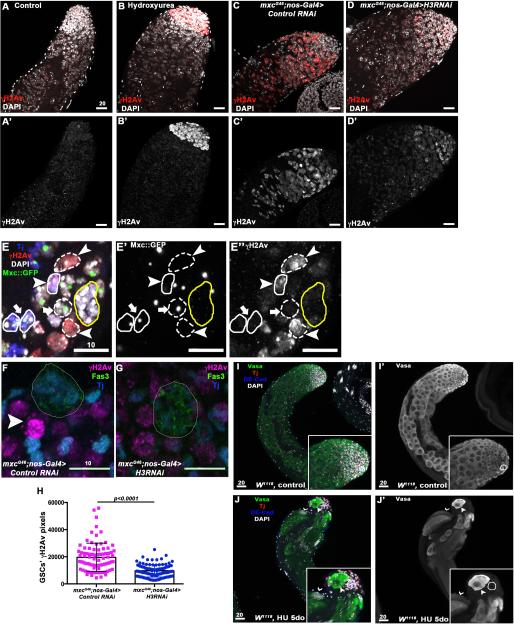
Figure 4. Persistent replicative stress leads to premature differentiation and germline loss.
A-B’. Little to no staining for γH2Av is observed in control testes (A-A’). Hydroxyurea treatment induces replicative stress in mitotic germ cells leading to accumulation of γH2Av (red) at the testis tip (B-B’). C-D’ Intense staining for γH2Av is observed in testes from mxcG46 males (C-C’), which is reduced upon H3 RNAi expression in early germ cells (D-D’). (E-E”) Clone induction in the testis using the null mutant allele of mxc, mxcG48. The Tj+ cell (blue) adjacent to the hub (yellow circle) circled by a solid line and indicated by an arrowhead represents a CySC mutant for mxc (as shown by the loss of Mxc ::GFP, E’) with high level of γH2Av in comparison to wild-type cyst cells (solid circle, arrow, E”). As expected, mxcG48 mutant germ cell clones (dashed line, arrowhead) also express high level of γH2Av compared to wild-type germ cells (dashed line, arrow). F-H. mxc mutant GSCs display saturation of γH2Av staining (magenta, F, arrowhead), which is less frequent and intense upon RNAi-medited reduction in H3 (G) Cyst cells (Tj+, blue); hub (Fas3+, green). The levels of γH2Av are significantly reduced in GSCs expressing H3 RNAi (H). I-J’ Prolonged exposure to hydroxyurea recapitulates mxc phenotypes. (I-I”) Testes from control flies contain early germ cells (Vasa+, green) at the tip (hub is circled, DE-Cad+, blue) followed by growing spermatocytes. (J-J’) Wild-type flies fed hydroxyurea for 5 consecutive days are depleted for early germ cells and contain cysts with fewer than 16 cells (arrowhead) close to the hub (circled), indicating incomplete TA divisions and precocious differentiation. Germ cells undergoing terminal differentiation are also present at the tip of the testes, as shown by cysts of elongating spermatids (thin arrowhead). Scale bars are in μM.

