
Fig. 2.
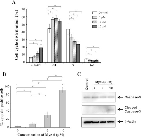
Myc-6-dependent induction of apoptosis. (A) Tali image-based cytometric analysis. MG63 cells were treated with the indicated concentrations of Myc-6 or left untreated. Following 72 h of Myc-6 exposure, the adherent and attached cells were collected and processed for Tali image-based cytometric analysis. Differences versus cells treated with or without Myc-6 were considered significant at p < 0.05. ∗p < 0.0001. The columns represent means ± SD. (B) Apopxin violet staining. Cells were treated as in (A). Forty-eight hours after the treatment, cells were incubated with apopxin violet 500 solution for 30 min, and then number of apopxin-positive cells was counted. ∗p < 0.01. (C) Cleavage of pro-apoptotic caspase-3. Cells were treated as in (A). Forty-eight hours after the treatment, cell lysates were prepared and analyzed by immunoblotting with the indicated antibodies.