
Fig. 3.
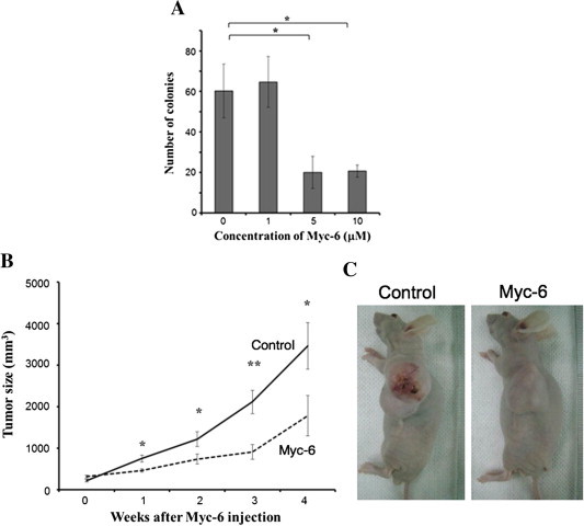
Tumor-suppressive activity of Myc-6 in vivo. (A) Anchorage-independent growth of MG63 cells. Cells were plated onto soft agar medium containing the indicated concentrations of Myc-6. Three weeks after Myc-6 treatment, number of colonies with a diameter of >100 μM was scored. Differences were considered significant at p < 0.05. ∗p < 0.01. The columns represent means ± SD. (B and C) Immuno-deficient nude mice bearing subcutaneously developed tumors derived from MG63 cells were injected intravenously with or without Myc-6 (6 μg/kg) once a week. At the indicated time periods after the first injection, tumor volume was measured. p < 0.05 was considered statistically significant. ∗p < 0.05; ∗∗p < 0.01 (B). Representative photographs of tumors were taken 4 weeks after Myc-6 injection (C).