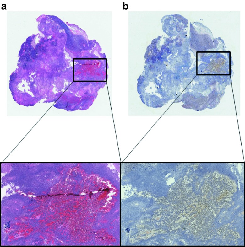
Figure 6.
Histological analyses of treated mouse tissues. Tumors and organs (liver, spleen, kidney, and brain) from the various groups were harvested at necropsy. (a) Immunohistochemistry specific for VSV was performed on sectioned tissues. Positive staining was observed only in combination treated tumors and can be visualized (brown) in the expanded magnification. (b) Consecutive sections were stained with H&E for general morphology and confirm the denucleation and tumor cell destruction caused by the combination therapy.

