
Figure 6.
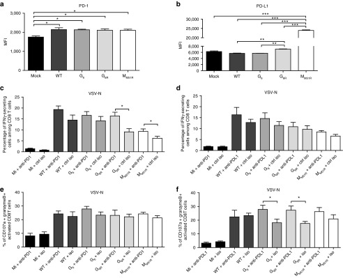
Expression of PD-1 and PD-L1 on B16gp33 cells following vesicular stomatitis virus (VSV) infection. B16gp33 cells were infected at a multiplicity of infection of 10 or mock infected. At 24 hours postinfection, cells were labeled to measure (a) PD-1 and (b) PD-L1 expression by flow cytometry. Data are the mean ± SEM and representative of two independent experiments in triplicates. B16gp33-bearing mice (n = 6–9) were also infected locally at the tumor site with 5.0 × 10⁸ PFU of WT or mutant VSV on day 7, 9, and 11 and treated either with (c,e) anti-PD-1 (250 μg), (d,f) anti-PD-L1 (200 μg) or isotype control antibodies on day 6, 10, and 14. On day 8 following the first VSV dose, splenocytes were isolated and stimulated ex vivo with VSV-N (RGYVYQGL) to analyze cytokines secretion. Data show the combination of three independent experiments. *P < 0.05, **P < 0.01, ***P < 0.001.