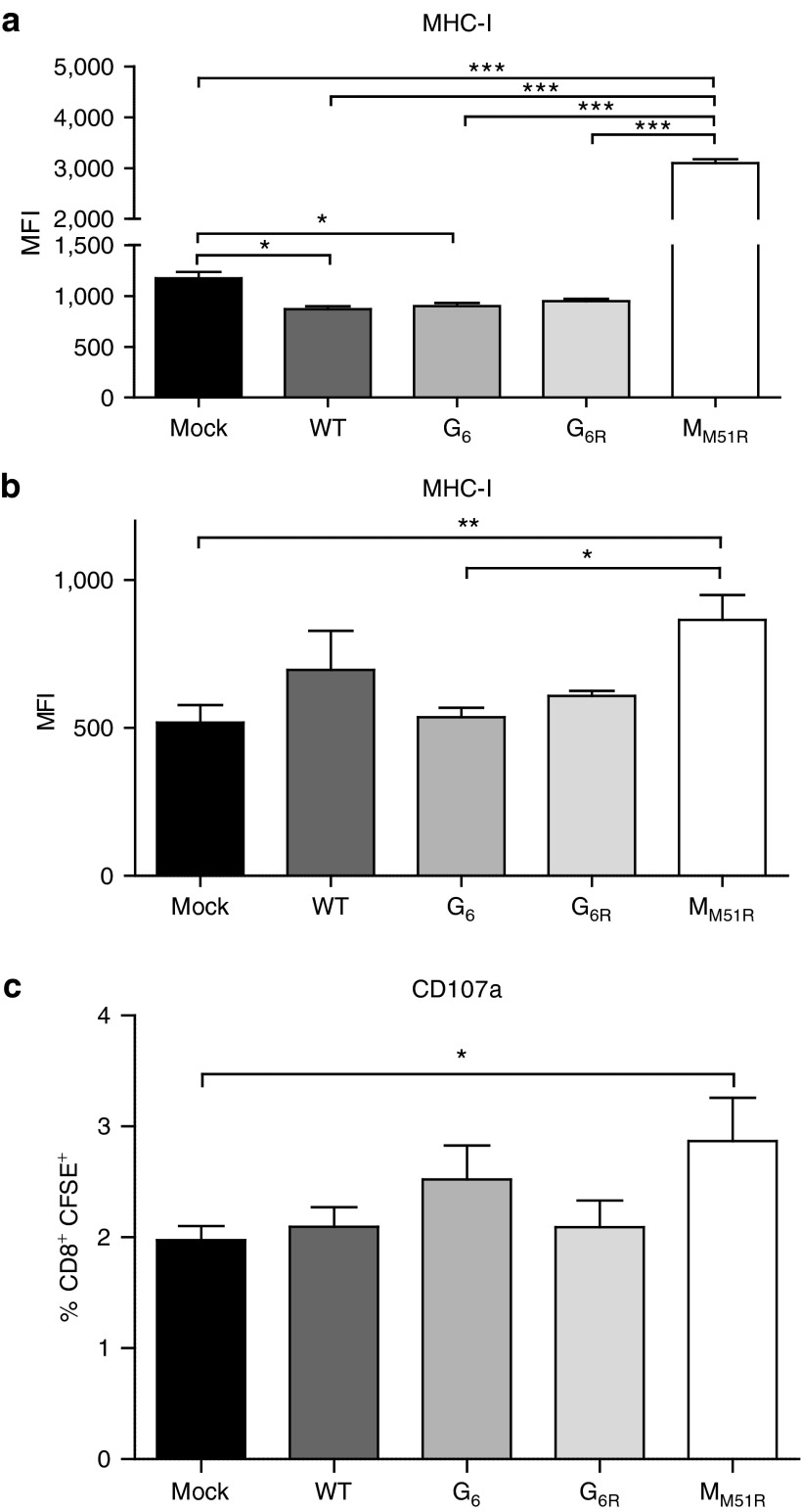
Figure 7.
Expression of major histocompatibility complex class-I (MHC-I) on B16gp33 cells following vesicular stomatitis virus (VSV) infection. (a) B16gp33 cells were infected at an multiplicity of infection of 10 or mock-infected. At 24 hours postinfection, cells were labeled to measure MHC-I expression by flow cytometry. Data are the mean ± SEM and representative of two independent experiments in triplicates. (b) B16gp33-bearing mice (n = 4–5) were infected locally at the tumor site with 5.0 × 10⁸ PFU of WT or mutant VSV on day 7. The following day, tumors were isolated and (b) labeled for the detection of MHC-I expression on CD45− cells or (c) cocultured with CFSE-labeled P14 transgenic splenocytes to analyze degranulation of CD8+CFSE+ cells. *P < 0.05, **P < 0.01, ***P < 0.001, CFSE, carboxyfluorescein succinimidyl ester.

