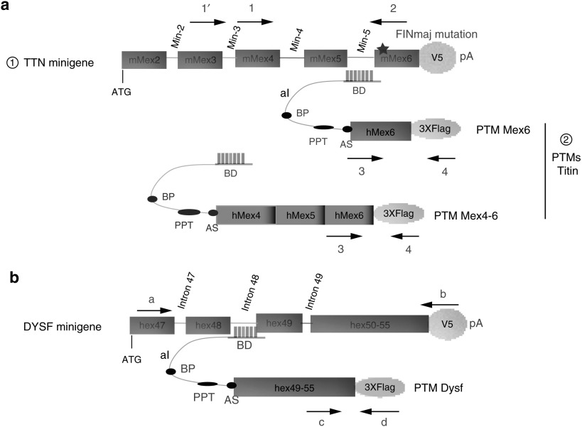
Figure 1.
Trans-splicing strategy of titin and dysferlin minigenes. (a) Diagram of the trans-splicing tools used for the 3′ titin exon(s) replacement (please note that the diagram is not drawn to scale). From top to bottom are depicted: : The mouse titin minigene construct used for the experiments in which the last 5 exons and last 4 introns of the mouse TTN gene were fused to a V5 epitope followed by a polyadenylation (pA) signal from SV40. The star symbolizes the presence of the FINmaj mutation. : The titin PTMs Mex6 and Mex4-6 consisting of a binding domain (BD) targeting the titin Min-5 or Min-3 introns, followed by an artificial intron (aI) containing a branching point (BP), a polypyrimidine tract (PPT) and an acceptor site (AS) followed by the WT last or last three exons of human titin fused to a 3xFlag epitope. The location of the oligonucleotides used to discriminate the trans-spliced RNA from the minigene and PTM transcripts is indicated by arrows 1-1′-2–3 and 4. Their sequences are given in Supplementary Table S1. (b) Diagram of the trans-splicing tools used for 3′ dysferlin exon replacement. The minigene carries the genomic sequence from exon 47 to intron 49, followed by a merge of the last 6 dysferlin exons fused to a V5 tag followed by a SV40 pA signal. The PTM provides the last 6 exons of dysferlin and carries the same aI as the titin PTM, with a specific BD that targets dysferlin intron 49. The location of the oligonucleotides used to discriminate trans-spliced RNA is indicated by arrows a, b, c, and d, and their sequences are given in Supplementary Table S1.

