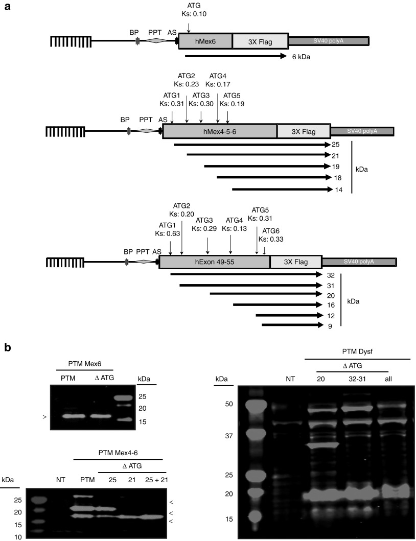
Figure 5.
Confirmation of translation from an open reading frame (ORF) present on the pre-trans-splicing molecule (PTM). (a) Location of the ORF relative to the PTM sequences. Each ORF with a significant Kozak score as defined by ATGPR software is indicated by an arrow below the diagram of each PTM. The position of the ATG, the Kozak score (Ks), and the corresponding predicted polypeptide weight is also indicated. AS, acceptor site; BP, branching point; PPT, polypyrimidine tract. (b) Analysis of the consequences of mutation of the ATG in the PTM. Left panel (top): western blot (WB) performed on HER911 cells transfected by titin PTM Mex6 or PTM Mex6 ΔATG. This experiment demonstrates that the observed 16 kDa protein is not translated from the corresponding ATG. Left panel (bottom): WB performed on HER911 cells transfected by titin PTM Mex4-6 or the PTM depleted of a different ORF initiating ATG (ΔATG25 or 21, or both 25+21). These ATG mutations abolished the proteins at 25 or 21 kDa or both, respectively. Right panel: WB performed on HER911 cells transfected by PTM Dysf or PTM Dysf depleted of the first two ATG (ΔATG32+31), the third ATG (ΔATG20), and finally, the ATG32, 31, 20, and the fourth ATG (ΔATGall). Disruption of ATG32 and 31 led to the disappearance of the 32 and 31 kDa proteins, respectively. ATG20 disruption does not show any protein disappearance on WB. The ATG16 disruption leads to the disappearance of the 16 kDa protein.

