Figure 1.
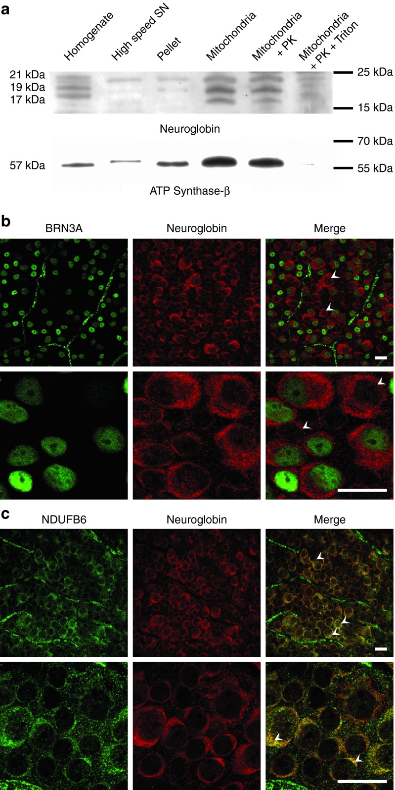
Neuroglobin (NGB) subcellular localization and distribution in mouse retinas. (a) Western blot detection of NGB and ATP synthase-β proteins in the different subcellular fractions from mouse retinas. Homogenates, high-speed supernatant obtained after mitochondrial centrifugation, pellet containing nuclei and unbroken cells, and mitochondrial fractions were subjected to western blot analysis after electrophoresis of 30 μg per sample. To confirm the presence of the NGB inside the mitochondria, the enriched mitochondrial fractions were treated with 150 µg/ml of proteinase K (PK) in the presence or absence of 1% Triton X100 at 4 °C for 30 minutes. The western blots were examined with different antibodies (Supplementary Table S1). The “PageRuler Plus Prestained Protein Ladder” allowed the estimation of apparent molecular mass of each signal; in the right margin is annotated the markers with similar electrophoretic properties. (b) Immunofluorescence analysis of retinal flat mounts from adult mice immunostained for NGB and BRN3A proteins. BRN3A labeling was exclusively nuclear (green). The prominent NGB labeling (red) was punctuated in the cytoplasm. Some NGB-positive cells did not show BRN3A labeling, they might be displaced amacrine cells or astrocytes which reside in the ganglion cell layer (white arrowheads). The scale bar represents 50 µm for all panels. (c) Retinal flat mounts from adult mice were double labeled for NGB (red) and the mitochondrial protein NDUFB6, a respiratory chain complex I subunit (green). The yellow-orange pixels show that NGB and NDUFB6 were in close apposition within numerous cells (white arrowheads). The scale bar represents 50 µm for all panels.

