Figure 7.
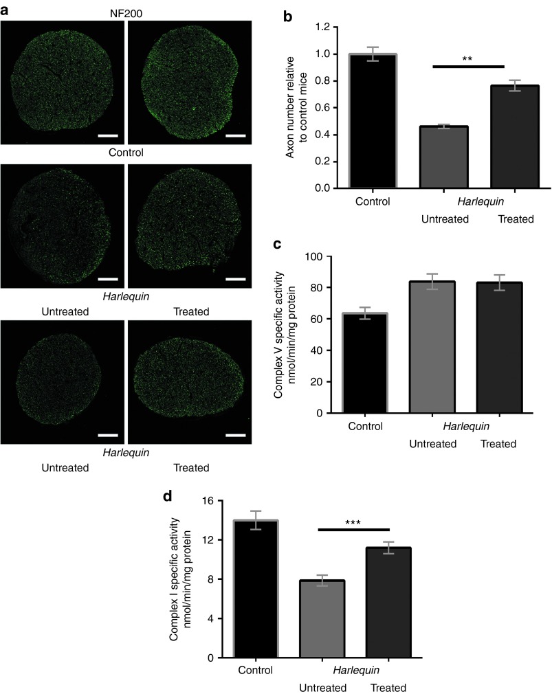
Morphological and functional evaluation of optic nerves from Harlequin mice after ocular AAV2/2-Neuroglobin treatment. (a) Independent proximal optic nerve transversal sections (near the globe) from 7-month-old control and Hq mice were immunolabeled with antibodies against the heavy chain (200 kDa) subunit of neurofilaments (NF200, green). The scale bars are equivalent to 50 µm. (b) Bar graph of axon number in controls and Hq optic nerves; six animals per group were evaluated after immunolabeling for NF200, and two independent transversal sections were entirely counted for positive spots using the ImageJ software. Results are illustrated relative to the axon number in optic nerves from aged-matched controls. The axon number per optic nerve in Hq-untreated mice was significantly different relative to controls or Hq subjected to AAV2/2-NGB intravitreal injection (P = 0.0022). (c,d) Specific complex V (CV) and complex I (CI) enzymatic activities were assessed in single optic nerves isolated from mice aged about 7–8 months: 38 controls and 29 Hq mice in which one eye was subjected to AAV2/2-NGB intravitreal injection and the contralateral one remained untreated. The successive measurements of CI and CV activities were expressed as nanomoles of oxidized NADH/min/mg protein. Histograms illustrate CV activity (b) and CI (c) as mean ± SEM of each assay per optic nerve measured in triplicate.

