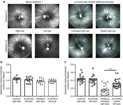
Figure 8.

Preservation of nerve fibers in AAV2/2-neuroglobin-treated eyes protects Harlequin mouse vision. (a) Confocal scanning laser ophthalmoscope fundus imaging of two Hq mice: images were collected in 4-week-old animals before AAV2/2-NGB administration, which was performed in their left eyes (left panel). Animals were evaluated monthly thereafter; images shown in the right panel were taken 6 months postinjection. Yellow discontinued lines show retinal regions with obvious nerve fiber loss in untreated eyes; for mouse # 1, the inferior region was the most affected, while for mouse # 2, the temporal area displays few RGC axons. Eyes treated with AAV2/2-NGB exhibited a marked preservation of optic fibers in all the retinal areas examined. Out of the 45 Hq mice subjected to AAV2/2 intravitreal injection in their left eyes and extensively evaluated with SLO, more than 50% exhibited a prevention of RGC axonal damage. (b,c) The Optomotry set-up allowed the determination of the optokinetic tracking threshold measurements (cycles per degree) for left and right eyes, independently scored (clockwise and counterclockwise responses respectively) under photopic conditions. Right and left eye sensitivities) for Hq (n = 14) and control (n = 14) mice aged 6–8 weeks are illustrated in (b) as means ± SEM of measures performed 2–3 times 4–6 days apart. (c) Histogram shows right and left eye sensitivities (Optokinetic response thresholds) for 7–8-month-old control mice (n = 25) and Hq mice subjected to AAV2/2-NGB injection in their left eyes (n = 25). Hq mice were evaluated at 3 and 6–7 months postinjection (the test was performed three times each time, 2–5 days apart), values presented are the means ± SEM of measures performed 6–7 months after AAV2/2-NGB administration and before euthanasia.
