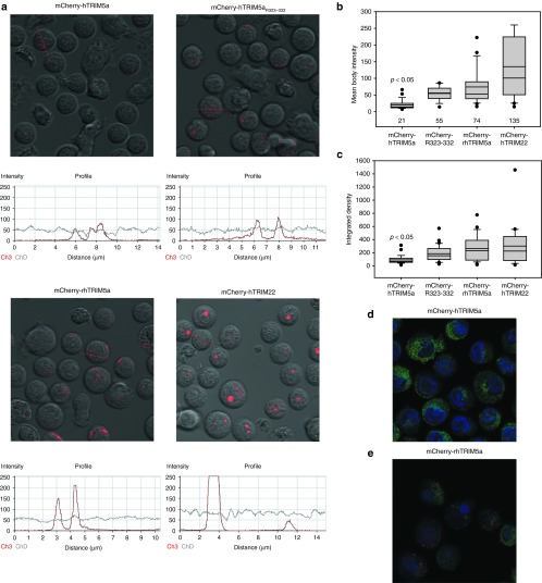
Figure 1.
The ability to form distinct and pronounced cytoplasmic bodies correlates with TRIM5α restriction activity. (a) Primary human CD4 T cells were activated with CD3/28 coated beads and transduced with the indicated mCherry-TRIM5α fusion or mCherry-TRIM22. Confocal microscopy was used to visualize and quantify the intensity of mCherry-TRIM5α cytoplasmic bodies in live human CD4 T cells on day 8 post-transduction. (b) Summary and quantification of the mean body intensity of cytoplasmic bodies in all cells shown in Figure 1a, as determined using the Zeiss ZLM Image Browser software (Carl Zeiss Microscopy LLC, Thornwood, NY). (c) Summary and quantification of the integrated density of cytoplasmic bodies in all cells shown in Figure 1a via ImageJ (http://fiji.sc/). For each condition, 15–20 cells and over 100 cytoplasmic bodies were analyzed per panel. Statistical testing to determine whether huTRIM5α formed less pronounced cytoplasmic bodies than other TRIM5α isoforms was performed using the Kruskal–Wallis one-way analysis of variance on Ranks test with Dunn's Method to adjust for multiple comparisons, utilizing Sigma Plot 11.0 software (Systat Software, Chicago, IL). (d) Imaging mCherry-huTRIM5α bodies in the cytoplasm (green) of fixed CD4 T cells with optimized exposure time; nucleus (blue) stained with DAPI. (e) Fixed cells expressing mCherry-rhTRIM5α bodies in the cytoplasm (green) with DAPI nuclear stain. DAPI, 4‘,6-diamidino-2-phenylindole.

