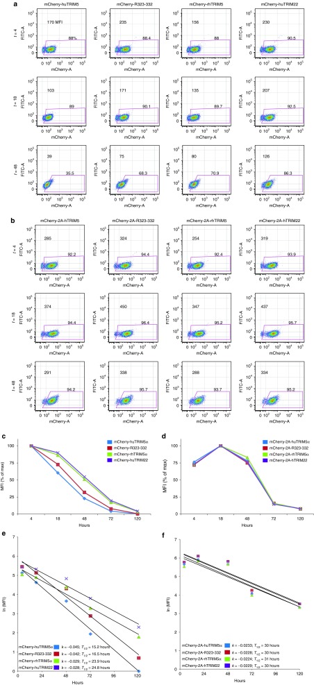
Figure 3.
HuTRIM5α is less stable than rhTRIM5α in primary human CD4 T cells. Resting primary human CD4 T cells were electroporated with 20 µg of in vitro transcribed RNA encoding mCherry-TRIM5α fusion proteins (a,c,e) or an mCherry-T2A-TRIM5α cassette (b,d,f), which expresses mCherry and TRIM in tandem but separately. (a,b) The mean fluorescence intensity (MFI) of mCherry was monitored by flow cytometry at T = 4, 18, 48, 72, and 120 hours after electroporation; CD4 T cells were CD3/28 costimulated at T = 18 hours. (c,d). Line graphs showing the decay of mCherry normalized to input values at T = 4 hours. (e,f) By taking the natural log of the MFI values and plotting them versus time, the slope or rate of decay (k), and half-life (T1/2) were calculated for each mCherry-TRIM5α fusion protein (e), and mCherry expressed separately but in an equimolar manner with TRIM5α or TRIM22 vis T2A motif (f), using the equation (T1/2 = ln(2)/k) (ref. 28). Results are representative of four independent experiments.

