Figure 1.
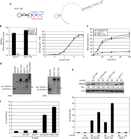
GL21.T-let conjugate specificity and processing. (a) Scheme (left panel) and secondary structure predicted by RNA structure v4.5 (right panel, free energy: −44.5 J/mol) of GL21.T-let. Guide strand is in red. (b) Left panel: binding of 50 nmol/l radiolabeled GL21.T or GL21.T-let on A549 (Axl+) and MCF-7 (Axl−) cells. Right panel: binding isotherm of GL21.T-let to soluble extracellular domain of Axl (EC-Axl). (c) Time-dependent internalization of radiolabeled GL21.T, GL21.T-let, or a scrambled sequence of GL21.T (scraGL21.T) used as negative control. Results are expressed as percent of internalized RNA relative to total bound. (d) Left panel: the indicated RNAs were untreated or treated with recombinant Dicer, resolved on a nondenaturing polyacrylamide gel and stained with ethidium bromide. Right panel: GL21.T-let containing radiolabeled guide strand was incubated with Dicer, resolved on gel, and analyzed by autoradiography. Size of single strand (ss) let-7g guide and expected size of miRNA duplex (ds) are indicated. (e) A549 (Axl+) cells were transfected with let-7g miRNA mimic (let-7g-miR), control-microRNA (ctrl-miR), GL21.T, or GL21.T-let. HMGA2 protein was analyzed by immunoblotting (upper panel) or let-7g miRNA was measured by RT-qPCR (lower panel), 48 hours after transfection. Values below the blots indicate signal levels relative to mock-treated cells (indicated as “-”), arbitrarily set to 1 (with asterisk). Intensity of bands was calculated using ImageJ (v1.46r). Equal loading was confirmed by immunoblot with anti-α-tubulin (αTub) antibodies. (f) RNAs from A549 (Axl+) cells transfected with let-7g-miR, ctrl-miR, GL21.T-let, or GL21.T were immuprecipitated with anti-Ago2 antibody and processed for let-7g miRNA RT-qPCR. No let-7g miRNA was immunoprecipitated with negative control mouse IgG (data not shown). In (b, e, and f) error bars depict mean ± SD (n = 3). HMGA2, high mobility group AT-hook 2; miRNA, microRNA; RT-qPCR, reverse transcription quantitative polymerase chain reaction.

